 |
|
|
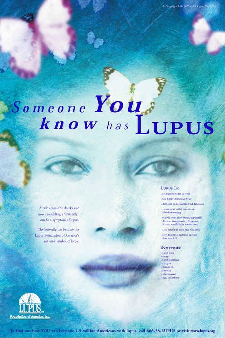
Early signs of lupus, even before a diagnosis can be made? Friday, October 26, 2007 Clinical criteria for systemic lupus erythematosus precede diagnosis and associated autoantibodies are present before clinical symptomsWhat is the topic? Diagnosing lupus is can sometimes be difficult. Because lupus symptoms may come and go and resemble other illnesses, it may take months or even years before a doctor can sort out what is going on. During this run-up period, most doctors agree, lupus disease activity is probably taking place, and lupus-related antibodies may be present even before symptoms occur. What did the researchers hope to learn? The researchers wanted to find out which symptoms tended to appear early on in most lupus patients and what antibodies might be present before certain clinical symptoms appear. They hoped this kind of analysis might provide clues about lupus disease activity and what course the disease might take even before symptoms are apparent. Who was studied? The researchers selected 130 patients who were diagnosed with lupus while they were in the United States military. Whenever someone joins the military, he or she has a baseline medical examination that includes an in-depth physical examination, medical history, and blood tests. The researchers would be able to use that information to look for signs of lupus and run tests for antibodies that were present before the diagnosis was made. How was the study conducted? Medical records for each of the 130 patients in the study were reviewed, and the researchers collected information about various features of lupus if and when they occurred, and if they appeared before the lupus diagnosis was made, how many months/years before. They also ran antibody tests on the blood samples to see what antibodies might have been present before clinical symptoms appeared. The researchers compared all of this information for patients based on their ethnic background, age when they were diagnosed with lupus, and the time frame separating symptoms’ appearance and diagnosis. What did the researchers find? Of the 130 patients studied, 104 had at least one clinical feature present before the diagnosis was made. Most of these patients (74) had only a single feature. The initial feature varied widely from patient to patient, but the most common early symptom was arthritis. In terms of time, discoid rash and seizures tended to be the earliest findings, occurring nearly two years before the diagnosis was made; interestingly, central nervous system involvement, which was one of the earliest problems to appear in patients in the study, did not commonly appear after the diagnosis. There were also interesting differences across populations. Men were more likely than women to have kidney disease as a first symptom, and Caucasians more likely than African Americans to have early rash on the cheeks and rashes that were made worse in the sun. Of the 104 patients who had clinical symptoms before their diagnosis, antibodies were often detectable before these symptoms. Antinuclear antibodies (ANA) showed up in 81 before the initial symptom. Seventeen patients had rheumatoid factor present when their blood was tested; arthritis developed later in 16 of them. As for anti-dsDNA and anti-C1q antibodies, which have been associated with kidney disease in lupus, 80 patients were positive for anti-dsDNA, and 38 of these patients had kidney disease; in 92 percent of these cases, the anti-dsDNA antibodies were detected prior to or at the same time as the diagnosis of kidney disease. About half of the patients who had anti-C1q antibodies developed kidney disease, but half did not; however, among the 18 anti-C1q patients who did have kidney disease, in 13 cases the antibodies showed up before the kidney disease was diagnosed, on average nearly one and a half years beforehand. What were the limitations of the study? The study was limited by the fact that the researchers had to rely on previously collected data, which had been provided for other purposes rather than specifically to look at lupus symptoms and lupus disease. Therefore, some lupus symptoms (like mouth sores) might not have been recorded because they weren’t really being looked for and they weren’t considered important in the overall health record of the patient at the time. Since the doctors following these patients before they were known to have lupus would not usually have been rheumatologists, there might have been many features of lupus that were simply missed. Also, though the researchers had at least one pre-diagnosis blood sample for each patient in the study, for some that was all they had, and the time intervals between that blood test and the lupus diagnosis varied greatly. For some patients there may have been changes in the antibody profile during that interval that the researchers wouldn’t have known about. What do the results mean for you? This study reinforces the notion that a lot of lupus disease activity may be taking place without patients’ or their doctors’ knowing it, in many cases long before the patient is diagnosed with lupus. However, the presence of certain antibodies may be a sign that later clinical symptoms are likely to appear, and so finding these antibodies early on when a patient has only one or a couple of symptoms may help doctors decide how they manage different patients, both before and after they have enough clinical criteria to determine a patient has lupus. On the other hand, it is also known that many family members of patients with lupus have lupus-related antibodies for many years without ever developing lupus. Some people can have these antibodies detectable temporarily during a viral illness or for unknown reasons, and then they go away. And as people age, they sometimes develop lupus-like antibodies as well, without becoming ill. The development of certain autoantibodies is probably one of several events that have to occur before a person develops lupus.
~~~ New symptom related to neonatal lupus What is the topic? Neonatal lupus erythematosus (NLE) is a rare but serious condition that can occur in newborn babies, and is related to anti-Ro (SSA) and/or anti-La (SSB) antibodies, which can cross the placenta in pregnancy from the mother to the fetus. A number of symptoms are seen in infants with NLE, most commonly a skin rash or liver involvement, both of which go away over time as the infant’s own immune system replaces the mothers circulating antibodies. However, a potentially serious heart condition called congenital heart block also can develop and will require medical attention. Developing babies of women with anti-Ro antibodies need to be monitored in the womb for heart block, though only a small number of pregnancies in women with lupus will result in this serious complication. In addition, some reports point to neurological symptoms that may be present in rare cases of NLE. What did the researchers hope to learn? The researchers sought to determine if infants born with NLE were at greater risk for hydrocephalus, a condition characterized by excess spinal fluid in or around the brain, which in turn contributed to macrocephaly, an enlarged head size. Who was studied? The study followed 87 infants born to selected, high risk women with anti-Ro antibodies and who were seen at the Hospital for Sick Children (HSC) in Toronto. Of the 87 infants studied, 47 were diagnosed as having NLE. How was the study conducted? Each of the 87 infants enrolled in the study was seen at least once in a follow-up visit, and more than 90% were seen more than once. What did the researchers find? The researchers found that the infants born to anti-Ro positive mothers developed hydrocephalus and macrocephaly at higher rates than would be expected. The rate of hydrocephalus was higher in both the group of infants with NLE and those identified as otherwise healthy. The researchers suggest that hydrocephalus be considered a new and independent manifestation of NLE, which may occur in association with other symptoms of NLE or alone. It is also important to point out that four of the five NLE infants who developed hydrocephalus were neurologically healthy, and in all but one of the infants, the abnormalities in head size and fluid volume resolved spontaneously over the course of time after their second birthday. What were the limitations of the study? The results of this study were similar to other investigations that showed an association of macrocephaly with NLE. It would have been better to compare this outcome to babies born to other lupus patients without anti-Ro antibodies and to the babies born to a group of healthy mothers attending this same hospital. Specialist hospitals such as this one could be attracting an overall more high risk group of patients with or without the Ro antibodies. What do the results mean for you? Women with lupus who have anti-Ro antibodies who are pregnant or intend to become pregnant need to be aware of the possibility their baby may develop NLE, which can have serious complications, albeit the serious complications are rare. These women should receive specialist high risk prenatal care and their babies should be monitored after birth by knowledgeable pediatricians.
~~~ Antidepressant Use Linked to Bone Loss Thursday, October 25, 2007 Older men and women who take the most widely used type of antidepressant medication may be at increased risk for bone loss, according to the results of 2 large studies.Antidepressants known as selective serotonin reuptake inhibitors (SSRIs) are often the first line of defense in treating depression, a condition increasingly diagnosed in the elderly. SSRIs help to alleviate depression by blocking the action of proteins in the brain known as serotonin transporters. In recent years, scientists have discovered that serotonin transporters are also present in bone cells. Animal and clinical studies suggest that the protein plays an as-yet unknown role in bone health. To see how SSRIs and other types of antidepressants might affect bone density in older patients, researchers drew on data collected for 2 large ongoing studies of osteoporosis-related bone fractures in people age 65 and older. The studies—one focusing on women and the other on men—are jointly funded by NIH’s National Institute on Aging (NIA) and National Institute of Arthritis and Musculoskeletal and Skin Diseases (NIAMS). The findings from both studies were published in separate papers in the June 25, 2007, issue of the Archives of Internal Medicine. For the women’s study—led by Dr. Susan J. Diem at the University of Minnesota—the researchers evaluated bone mineral density in participants' hip bones over a 5-year period. Of the 2,722 women under study, about 7% were taking SSRIs, 4% were taking tricyclic antidepressants and the rest were taking neither. After adjusting for other factors—such as the severity of depression and use of calcium supplements—the researchers found that bone mineral density in SSRI users dropped by 0.82% each year, compared to an annual loss of 0.47% among those taking either tricyclic antidepressants or no antidepressants. In the other study—led by Dr. Elizabeth Haney at the Oregon Health and Science University—the scientists looked at bone density in the hips and the spines of 5,995 men. About 3% of the men were taking SSRIs, and about 3% were taking other antidepressants. Bone mineral density among SSRI users was 3.9% lower at the hip and 5.6% lower in the spine than in the other participants. Although the researchers linked SSRIs to bone loss, they stress that this work doesn’t prove that SSRIs are responsible. Their findings need to be confirmed by additional studies, and other possible contributing factors, like depression itself, need to be definitively ruled out. “Depression is a serious condition, and it’s important not to stop antidepressants abruptly or without talking to your physician,” Haney says. “However, it may be worth paying special attention to the things we know can prevent osteoporosis, such as exercise, taking adequate calcium and vitamin D.”—by Vicki Contie Labels: antidepressant, bone, depression
~~~ |
.:Find Me:. If you interested in content, please contact the Writer .:Want to Joint ?:. If you want to know more about lupus surferer's activities and want to donor your help and money, go here Need more consult ?, go here .:acquaintances:.
The Enterprise .:New Book:. .:talk about it:.
.:archives:.
.:Link-link website Lupus:.
Lupus Org .:credits:.
|